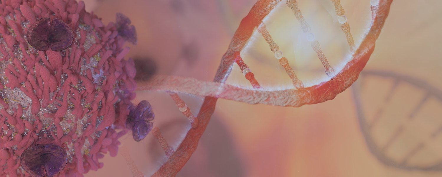
Pathology and Biorepository Core
Exceptional Service. Rigorous Science.
VAI’s Pathology and Biorepository Core integrates anatomic pathology expertise with biorepository and biospecimen science for investigator- and project-driven research. The Core builds upon strengths in standard histology, microscopy and biobanking, along with novel technologies to test and apply best practices in biospecimen science. The Core’s focus on pathology and molecular quality metrics provide complementary emphasis on high-quality biospecimens and interpretable results with which to validate experimental models and extend them to clinical samples, thereby advancing the Institute’s mission to improve the health and enhance the lives of current and future generations.
VAI’s Biorepository is nationally and internationally recognized, once serving as the NCI Comprehensive Biospecimen Resource for the Cancer Human Biobank (caHUB), the Biospecimen Pre-analytical Variables Program and the Genomic Tissue Expression (GTEx) project, and presently as the Biospecimen Core Resource for the National Cancer Institute’s Clinical Proteomic Tumor Analysis Consortium (CPTAC) and the National Cancer Institute’s Cancer Moonshot Program. It also provides biorepository services for the Multiple Myeloma Research Foundation and the Tuberous Sclerosis Alliance.
VAI’s Biorepository has been accredited by the College of American Pathologists (CAP) since 2012. Its accreditation number is 8017856.

Acknowledgment in Publications
Authors who publish articles containing work performed by VAI’s Core Technologies and Services are asked to follow these guidelines in their acknowledgements:
- Acknowledge the contributions of VAI’s Pathology and Biorepository Core by name in the manuscript.
- Consider authorship for more significant intellectual and planning contributions.
- Notify Core Services staff of successful publications and awarded grant applications. This assists in tracking and maintaining institutional records, which help inform the Cores’ quality services.
OUR IMPACT
We’re raising thousands to save millions.
We’re turning hope into action for the millions of people around the world affected by diseases like cancer and Parkinson’s. Find out how you can help us make a difference.
- 121 peer-reviewed papers published in 2023
- 62 peer-reviewed papers published in high-impact journals in 2023
- 55 clinical trials launched to date
Services
VAI’s Pathology and Biorepository Core offers a comprehensive range of biospecimen services, including histology, pathology and biospecimen processing and storage. The Core manages service requests through Agilent CrossLab, which provides service descriptions as well as options to schedule equipment and request services. Pricing also is available in CrossLab (See Schedule Equipment and/or Request Services). Current CrossLab users can login here.
VAI staff that need a login (or further CrossLab support) should contact Lori Moon. MSU and external users that need a CrossLab account/login should go here for account creation. After logging in, external users should click “list all cores” under the Core Facilities section in the left menu and scroll down to “Cores at Van Andel Institute.”
Biorepository Molecular Processing HistopathologyVAI has a FISMA ATO rating of low for present National Cancer Institute projects, meaning that the Institute has extensive systems for monitoring, backup and security of sensitive and important data. The Biorepository uses the Biological Specimen Inventory (BSI) database and software, developed by NCI and IMS for the management of biospecimens. VARI’s entire Core Technologies and Services team use Agilent CrossLab (formerly iLab) software to order and track specimens through production and billing.
Agilent Bioanalyzer 2100
Agilent TapeStation
Aperio Scanscope AT2
Covaris cryoPREP CP02
CryoXtract CXT 353
Dako Autostainer Link 48
Leica Cryostats
Leica Embedding Center
Leica Microtomes
Miltenyi autoMACS Pro Separators
MVE CryoCarts
NanoDrop 8000
Pathology Devices TMA Arrayer
Promega Maxwell RSC
Qiagen TissueLyzer II
Qubit Fluorometers
Sakura Tissue-Tek Prisma Plus
Sakura Tissue-Tek VIP Tissue Processors
Thermo Scientific Shandon Cytospin 4
-80°C and -20°C mechanical freezers, laboratory-grade refrigerators
MVE Liquid Nitrogen (LN2) freezers
VAI’s Biorepository provides services for a number of large-scale national and international projects. For more information click a project below or please contact us at [email protected].
Biospecimen Pre-Analytical Variables (BPV)
Cancer Moonshot Biobank
Clinical Proteomic Tumor Analysis Consortium (CPTAC)
Genotype-Tissue Expression (GTEX) Project
Multiple Myeloma Research Foundation’s CoMMpass Study
TSC Alliance Biosample Repository
Van Andel Institute–Stand Up To Cancer Epigenetics Dream Team
FAQs
VAI does not have a program specifically for organ and body donation. Many hospitals and universities do have these donation programs; however, the process can be lengthy and costly. We recommend consulting those organizations as early as possible to discuss your options.
VAI’s Biorepository does accept tumor tissue samples according to specific guidelines. If you would like to donate tumor tissue to VAI, please consider discussing the option with your physician and ask him or her to reach out to Core Director Dan Rohrer. VAI has a sample collection agreement with Mercy Health St. Mary’s Health System in Grand Rapids and can work with other institutions to assess the best options for donation.
Please visit our Services section for specific information on each service.
Cost depends on a number of factors such as the sample type and preparation requested as well as additional processing requirements. This is best determined through the application process. For more information, please visit our Biorepository page.
VAI staff that need a login (or further CrossLab support) should contact Lori Moon. MSU and external users that need a CrossLab account/login should go here for account creation. After logging in, external users should click “list all cores” under the Core Facilities section in the left menu and scroll down to “Cores at Van Andel Institute.”
Please acknowledge us as:
“Van Andel Institute, Pathology and Biorepository Core, Grand Rapids, MI”
We store a variety of biospecimens including serum, plasma, saliva, cells, and tissues that are formalin-fixed paraffin-embedded (FFPE) and frozen. We also store their respective molecular analytes such as DNA and RNA or pulverized tissues for proteomics.
If you are a VAI investigator or fund a disease-specific biorepository, we would be interested in storing your biospecimens. To find out if this option fulfills your interest, please contact us at [email protected].

Ensuring the quality of biospecimens and biospecimen research are the central to the Pathology and Biorepository Core’s mission. To this end, the Core focuses on efforts to improve the management of biospecimens using evidence-based best practices and leverages the extensive experience of its director and staff in national efforts to harmonize common biorepository procedures and set standards. VAI’s Biorepository is accredited by the College of American Pathologists through its Biorepository Accreditation Program (no. 8017856).
The value of human biospecimens available for research dramatically increases when linked with their accumulated clinical and molecular profiling data. The Core inventories and links all human biospecimens with this data to provide high-quality biospecimens for research while maintaining human subjects protections. A complete Quality Management System (QMS) governs all aspects of the Core. The QMS is overseen by a manager and supported by quality assurance specialists. All of the Core’s processes follow standard operating procedures, which ensure a consistent and rigorous approach to sample handling and analysis. Key elements of the QMS are:
- Standard operating procedures (SOPs)
- Personnel training and competency
- Equipment maintenance and qualification
- Data management and record keeping
- Routine audits
- Reagent/material tracking and labeling
- CAPA and continuous process improvement
Quality Control Systems
The Core also utilizes quality control measures to assess specimen integrity and laboratory work. Examples of QC measures include:
- Inspection of specimen collection kits prior to distribution
- Inspection of all specimens received in the Core prior to assessioning or storage
- Research pathology review (daily and weekly QC slide review in addition to review of all tissue specimens prior to distribution)
- Positive and negative controls utilized during molecular and histological processing
- Quantification and qualification of molecular analytes (e.g., RIN values, spectrophometric measurements, fluorometric quantitation)
A centralized, in-house system for biospecimen tracking and molecular processing reduces costs through elimination of redundancies while also maximizing efficiency and economy of scale. Furthermore, the concerted and consistent application of best practices for molecular analyte generation facilitates downstream large-scale genomic and proteomic analyses, thereby maximizing the quantity and quality of data collected per sample.
Biorepository Storage Units
Temperature and humidity are controlled and monitored in each area of the facility by the building’s Tridium Control & Monitoring System, which includes dedicated air handlers to maintain temperature and humidity at 30–50 percent. Should humidity levels, temperature or other indicators fall outside (above or below) threshold limits, an alarm sounds in the security office and a system-generated message is simultaneously sent to designated Core staff and on-site security personnel for further action.
Emergency power is provided via two 1.5-megawatt on-site diesel generators for which a 10,000-gallon diesel fuel tank provides sufficient fuel to run. The generators ensure power for all vital facility operations, including lighting, HVAC, freezers, refrigerators, critical equipment and blowers on ventilated racks.
SELECTED PUBLICATIONS
Goralski TM, Meyerdirk L, Breton L, Brasseur L, Kurgat K, DeWeerd D, Turner L, Becker K, Adams M, Newhouse DJ, Henderson MX. 2024. Spatial transcriptomics reveals molecular dysfunction associated with cortical Lewy pathology. Nat Commun 15:2642.
Grit JL, Turner L, Essenburg CJ, Gallik KL, Dischinger PS, Shurlow ND, Pate MJ, Graveel CR, Steensma MR. 2024.Ex-vivo patient-derived explant model for neurofibromatosis type 1-related cutaneous neurofibromas. J Invest Dermatol 21:S0022-202X(24)00117-9.
Kitchen-Goosen SM, Schumacher H, Good J, Patterson AL, Boguslawski EA, West RA, Williams BO, Hostetter G, Agnew DW, Teixeira JM, Alberts AS. 2023. Endometrial hyperplasia with loss of APC in a novel population of Lyz2-expressing mouse in endometrial epithelial cells. Carcinogen 44(1):54–64.
Dues DJ, Tran Nguyen AP, Becker K, Ma J, Moore DJ. 2023. Hippocampal subfield vulnerability to α-synuclein pathology precedes neurodegeneration and cognitive dysfunction. npj Parkinsons Dis 9(125).
*Core included in acknowledgements
Cui W, Huang Z, Jin S-G, Johnson J, Lau KH, Hostetter G, Pfeifer GP. 2023. Deficiency of the Polycomb protein RYBP and TET methylcytosine oxidases promotes extensive CpG island hypermethylation and malignant transformation. Cancer Res.
Jang HJ, Hostetter G, MacFarlane AW, Madaj Z, Ross EA, Hinoue T, Kulchycki JR, Burgos RS, Tafseer M, Alpaugh RK, Schwebel CL, Kokate R, Geynisman DM, Zibelman MR, Ghatalia P, Nichols PW, Chung W, Madzo J, Hahn NM, Quinn DI, Issa JPJ, Topper MJ, Baylin SB, Shen H, Campbell KS, Jones PA, Plimack ER. 2023. A phase II trial of guadecitabine plus atezolizumab in metastatic urothelial carcinoma progressing after initial immune checkpoint inhibitor therapy. Clin Cancer Res: OF1–OF4.
O’Connell CL, Baer MR, Ørskov AD, Saini SK, Duong VH, Kropf P, Hansen JW, Tsao-Wei D, Jang HS, Emadi A, Holmberg-Thyden S, Cowland J, Brinker BT, Horwood K, Burgos R, Hostetter G, Youngblood BA, Hadrup SR, Issa J-P, Jones P, Baylin SB, Siddiqi I, Gronbaek K. 2022. Safety, outcomes and T cell characteristics in patients with relapsed or refractory MDS or CMML treated with atezolizumab in combination with guadecitabine. Clin Cancer Res 28(24):5306-5316.
Luchtefeld M, Jrebi N, Hostetter G, Osterholzer K, Dykema K, Khoo SK. 2022. Effect of doxycycline-release anastomotic augmentation ring on porcine colorectal anastomosis. J Surg Res 279:464–473.
Wilson MR, Skalski H, Reske JJ, Wegener M, Adams M, Hostetter G, Hoffman HH, Bernard JJ, Bae-Jump VL, Teixeira JM, Chandler RL. 2022. Obesity alters the mouse endometrial transcriptome in a cell context-dependent manner. Reprod Biol Endocrinol 20(1):163.
Li QK, Hu Y, Chen L, Schnaubelt M, Zhou DC, Li Y, Lu RJH, Thiagarajan, Mathangi, Hostetter G, Newton CJ, Jewell SD, Omenn G, Robles AI, Mesri M, Bathe OF, Zhang B, Ding L, Hruban RH, Chan DW, Zhang H. 2022. Neoplastic cell enrichment of tumor tissues using coring and laser microdissection for proteomic and genomic analyses of pancreatic ductal adenocarcinoma. Clin Proteom 19(1):36.
Yang CH*, Fagnocchi L*, Apostle S, Wegert V, Casani-Galdón S, Landgraf K, Panzeri I, Dror E, Heyne S, Wörpel T, Chandler DP, Lu D, Yang T, Gibbons E, Guerreiro R, Brás J, Thomasen M, Grunnert LG, Vaag AA, Gillberg L, Grundberg, E, Conesa A, Körner A, PERMUTE, Pospisilik JA. 2022. Independent phenotypic plasticity axes define distinct obesity subtypes. Nat Metab.
*Co-first authorship
**Highlighted in News & Views
***The Core contributed to this work
Bagchi A, Beddows I, Cornelius A, Robinson GW, Jewell SD. 2021. Rare cases of medulloblastoma with hypermutation. Cancer Rep e1521.
Bagchi A, Madaj Z, Engel KB, Guan P, Rohrer DC, Valley DR, Wolfrum E, Feenstra K, Roche N, Hostetter G, Moore HM, Jewell SD. 2021. Impact of preanalytical factors on the measurement of tumor tissue biomarkers using immunohistochemistry. J Histochem Cytochem 69(5)297–320.
GTEx Consortium. 2020. The GTEx Consortium atlas of genetic regulatory effects across human tissues. Science 369(6509):1318–1330.
*Part of special issue highlighting GTEx V8 data release. The Pathology and Biorepository Core serves as the Biospecimen Core Resource for GTEx.
Olivia M … Stranger BE. 2020. The impact of sex on gene expression across human tissues. Science 369(6509).
*Part of special issue highlighting GTEx V8 data release. The Pathology and Biorepository Core serves as the Biospecimen Core Resource for GTEx.
Kim-Hellmuth S … Lappalainen T. 2020. Cell type-specific genetic regulation of gene expression across human tissues. Science 369(6509).
*Part of special issue highlighting GTEx V8 data release. The Pathology and Biorepository Core serves as the Biospecimen Core Resource for GTEx.
Demanelis K … GTEx Consortium … Pierce BL. 2020. Determinants of telomere length across human tissues. Science 369(6509).
*Part of special issue highlighting GTEx V8 data release. The Pathology and Biorepository Core serves as the Biospecimen Core Resource for GTEx.
Ferraro NM …GTEx Consortium … Battle A. 2020. Transcriptomic signatures across human tissues identify functional rare genetic variation. Science 369(6509).
*Part of special issue highlighting GTEx V8 data release. The Pathology and Biorepository Core serves as the Biospecimen Core Resource for GTEx.
Mathieson W, Mommaerts K, Trouet JM, Mathay C, Guan P, Carithers LJ, Rohrer D, Valley DR, Blanski A, Jewell S, Moore HM, Betsou F. 2018. Cold ischemia score: An mRNA assay for the detection of extended cold ischemia in formalin-fixed, paraffin-embedded tissue. J Histochem Cytochem 18:22155418819967.
Vaseva AV, Blake DR, Gilbert TSK, Ng S, Hostetter G, Azam SH, Ozkan-Dagliyan I, Gautam P, Bryant KL, Pearce KH, Herring LE, Han H, Graves LM, Witkiewicz AK, Knudsen ES, Pecot CV, Rashid N, Houghton PJ, Wennerberg K, Cox AD, Der CJ. 2018. KRAS suppression-induced degradation of MYC is antagonized by a MEK5-ERK5 compensatory mechanism. Can Cell 34(5):807-822.
Mukherjee A, Patterson AL, George JW, Carpenter TJ, Madaj ZB, Hostetter G, Risinger JI, Teixeira JM. 2018. Nuclear PTEN localization contributes to DNA damage response in endometrial adenocarcinoma and could have a diagnostic benefit for therapeutic management of the disease. Mol Cancer Ther 17(9):1995–2003.
Manojlovic Z, Christofferson A, Liang WS, Aldrich J, Washington M, Wong S, Rohrer D, Jewell S, Kittles RA, Derome M, Auclair D, Craig DW, Keats J, Carpten JD. 2017. Comprehensive molecular profiling of 718 Multiple Myelomas reveals significant differences in mutation frequencies between African and European descent cases. PLoS Genet 13(11):e1007087.
Harlow ML, Maloney N, Roland J, Guillen Navarro MJ, Easton MK, Kitchen-Goosen SM, Boguslawski EA, Madaj ZB, Johnson BK, Bowman MJ, D’Incalci M, Winn ME, Turner L, Hostetter G, Galmarini CM, Aviles PM, Grohar PJ. 2016. Lurbinectedin inactivates the Ewing sarcoma oncoprotein EWS-FLI1 by redistributing it within the nucleus. Cancer Res 76(22):6657–6668.
Meng X, Vander Ark A, Lee P, Hostetter G, Bhowmick NA, Matrisian LM, Williams BO, Miranti CK, Li X. 2016. Myeloid-specific TGF-β signaling in bone promotes basic-FGF and breast cancer bone metastasis. Oncogene 35(18):2370-2377.
GTEx Consortium. 2015. Human genomics. The Genotype-Tissue Expression (GTEx) pilot analysis: multitissue gene regulation in humans. Science. 348(6235):648–660.
*Core contributions: Dana Valley, Dan Rohrer and Scott Jewell
Carithers LJ, Ardlie K, Barcus M, Branton PA, Britton A, Buia SA, Compton CC, DeLuca DS, Peter-Demchok J, Gelfand ET, Guan P, Korzeniewski GE, Lockhart NC, Rabiner CA, Rao AK, Robinson KL, Roche NV, Sawyer SJ, Segré A, Shive CE, Smith AM, Sobin LH, Undale AH, Valentino KM, Vaught J, Young TR, Moore HM, on behalf of the GTEx Consortium. 2015. A novel approach to high-quality post-mortem tissue procurement: The GTEx Program. Biopreserv Biobank 13(5):311–319.
*Core contributions: Dana Valley, Dan Rohrer and Scott Jewell
Keats J, Speyer G, Christofferson A, Stephenson K, Kurdoglu A, Russell M, Aldrich J, Legendre C, Cuyugan L, Adkins J, McDonald J, Helland A, Blanksi A, Hodges M, Rohrer D, Jagannath S, Siegel D, Vij R, Orloff G, Zimmerman T, Niesvizky R, Liles D, Fay J, Wolf J, Rifin R, Derome M, Auclair D, Liang W, Kim S, Guiterrez N, Kidd P, Jewell S, Craig D, Carpten J, Lonial S. 2015. Interim analysis of the MMRF CoMMpass Study: Comprehensive characterization of multiple myeloma patients at diagnosis reveals distinct molecular subtypes and clinical outcomes. Clin Lymphoma Myeloma Leuk 15:e44–e45.
Ensink E, Sinha J, Sinha A, Tang H, Calderone HM, Hostetter G, Winter J, Cherba D, Brand RE, Allen PJ, Sempere LF, Haab BB. 2015. Segment and fit thresholding: a new method for image analysis applied to microarray and immunofluorescence data. Anal Chem 87(19):9715-9721.
Valkenburg KC, Hostetter G, Williams BO. 2015. Concurrent Hepsin overexpression and adenomatous polyposis coli deletion causes invasive prostate carcinoma in mice. Prostate75)14):1579–1580.
MacKenzie TA, Schwartz GN, Calderone HM, Graveel CR, Winn ME, Hostetter G, Wells WA, Sempere LF. 2014. Stromal expression of miR-21 identifies high-risk group in triple-negative breast cancer. Am J Path 184(12):3217–3225.
Berger PL, Frank SB, Schulz VV, Nollet EA, Edick MJ, Holly B, Chang TA, Hostetter G, Kim S, Miranti CK. 2014. Transient induction of ING4 by MYC drives prostate epithelial cell differentiation and its disruption drives prostate tumorigenesis. Cancer Res 74(12): 3357–3368.
Robb JA, Gulley ML, Fitzgibbons PL, Kennedy MF, Cosentino LM, Washington K, Dash RC, Branton PA, Jewell SD, Lapham RL. 2014. A call to standardize preanalytic data elements for biospecimens. Arch Pathol Lab Med 138:526–537.
Hostetter G, Collins E, Varlan P, Edewaard E, Harbach PR, Hudson EA, Feenstra KJ, Turner LM, Berghuis BD, Resau JH, Jewell SD. 2014. Veterinary and human biobanking practices: enhancing molecular sample integrity. Vet Pathol 51(1): 270–280.
Lonsdale J, Thomas J, Salvatore M, Phillips R, Lo E, Shad S, Hasz R, Walters G, Garcia F, (including Jewell, SD) et al. 2013. The Genotype-Tissue Expression (GTEx) project. Nature Genetics 45(6): 580–585.
Jewell SD, Cordial CR. 2013. Silver staining of nucleolar organizer regions. J Histotech 19:241–256.
Spivak-Kroizman TR, Hostetter G, Posner R, Aziz M, Hu C, Demeure MJ, Von Hoff D, Hingorani SR, Palculict TB, Izzo J, Kiriakova GM, Abdelmelek M, Bartholomeusz G, James BP, Powis G. 2013. Hypoxia triggers hedgehog-mediated tumor-stromal interactions in pancreatic cancer. Cancer Res 73(11):3235-47.
Weiss GJ, Liang WS, Demeure MJ, Kiefer JA, Hostetter G, Izatt T, Sinari S, Christoforides A, Aldrich J, Kurdoglu A, Phillips L, Benson H, Reiman R, Baker A, Marsh V, Von Hoff DD, Carpten JD, Craig DW. 2013. A pilot study using next-generation sequencing in advanced cancers: feasibility and challenges. PLoS One 8(10):e76438.
Cao Y, Zhang ZL, Zhou M, Elson P, Rini B, Aydin H, Feenstra K, Tan MH, Berghuis B, Tabbey R, Resau JH, Zhou FJ, Teh BT, Qian CN. 2013. Pericyte coverage of differentiated vessels inside tumor vasculature is an independent unfavorable prognostic factor for patients with clear cell renal cell carcinoma. Cancer 119(2):313-324.
Resau, JH, Ho NT, Dykema K, Faber MS, Busik JV, Nickolov RZ, Furge KA, Paneth N, Jewell S, Khoo SK. 2012. Evaluation of sex-specific gene expression in archived dried blood spots (DBS). Int J Mol Sci 13(8):9599-9608.
Raz G, Allen KE, Kingsley C, Cherni I, Arora S, Watanabe A, Lorenzo CD, Edwards V DK, Sridhar S, Hostetter G, Weiss GJ. 2012. Hedgehog signaling pathway molecules and ALDH1A1 expression in early-stage non-small cell lung cancer. Lung Cancer 76(2):191-196.
Byron SA, Min E, Thal TS, Hostetter G, Watanabe AT, Azorsa DO, Little TH, Tapia C, Kim S. 2012. Negative regulation of NF-κB by the ING4 tumor suppressor in breast cancer. PLoS One. 7(10):e46823.
Weiss GJ, Liang WS, Izatt T, Arora S, Cherni I, Raju RN, Hostetter G, Kurdoglu A, Christoforides A, Sinari S, Baker AS, Metpally R, Tembe WD, Phillips L, Von Hoff DD, Craig DW, Carpten JD. 2012. Paired tumor and normal whole genome sequencing of metastatic olfactory neuroblastoma. PLoS One 7(5):e37029.
Asmann YW, Necela BM, Kalari KR, Hossain A, Baker TR, Carr JM, Davis C, Getz JE, Hostetter G, Li X, McLaughlin SA, Radisky DC, Schroth GP, Cunliffe HE, Perez EA, Thompson EA. 2012. Detection of redundant fusion transcripts as biomarkers or disease-specific therapeutic targets in breast cancer. Cancer Res 72(8):1921-1928.
Stephens B, Anthony SP, Han H, Kiefer J, Hostetter G, Barrett M, Von Hoff DD. 2012. Molecular characterization of a patient’s small cell carcinoma of the ovary of the hypercalcemic type. J Cancer. 3:58-66.
Diep CH, Zucker KM, Hostetter G, Watanabe A, Hu C, Munoz RM, Von Hoff DD, Han H. 2012. Down-regulation of Yes Associated Protein 1 expression reduces cell proliferation and clonogenicity of pancreatic cancer cells. PLoS One 7(3):e32783.
Edelman MJ, Hodgson L, Wang X, Christenson R, Jewell SD, Vokes E, Kratzke R. 2011. Serum vascular endothelial growth factor and COS-2/5-LOX inhibition in advanced non-small cell lung cancer: Cancer and Leukemia Group B 150304. J Thorac Oncol 6(11):1902–1906.
Moore HM, Kelly AB, Jewell SD, McShane LM, Clark DP, Greenspan R, Hayes DF, Hainaut P, Kim P, Mansfield E, Potapova O, Riegman P, Rubinstein Y, Seijo E, Somiari S, Watson P, Weier HU, Zhu C, Vaught J. 2011. Biospecimen reporting for improved study quality (BRISQ). J Proteome Res. 10(8):3429–3438.
Rimm DL, Nielsen TO, Jewell SD, Rohrer D, Broadwater G, Waldman F, Mitchell KA, Singh B, Tsongalis GJ, Frankel WL, Magliocco A, Lara JF, Hsi ED, Bleiweiss I, Badve SS, Chen B, Ravdin P, Schilsky R, Thor A, Berry DA. 2011. CALGB Pathology Committee guidelines for tissue microarray construction representing multi-center prospective clinical trial tissues for cancer and leukemia group B, Eastern Oncology Group, Southwest Oncology Group, and North Central Cancer Treatment Group. J Clin Oncol 29(16):2282–2290.
Savage SJ, Hostetter G. 2011. Genomic analysis by oligonucleotide array Comparative Genomic Hybridization utilizing formalin-fixed, paraffin-embedded tissues. Methods Mol Biol 700:185-98.
Bapat AA, Hostetter G, Von Hoff D, Han H. 2011. Perineural invasion and associated pain in pancreatic cancer. Nat Rev Cancer 11(10):695-707.
Whatcott CJ, Han H, Posner RG, Hostetter G, Von Hoff D. 2011. Targeting the tumor microenvironment in cancer: why hyaluronidase deserves a second look. Cancer Discov 1(4):291-296.
Harris CA, Resau JH, Hudson EA, West RA, Moon C, Black AD, McAllister JP. 2011. Effects of surface wettability, flow, and protein concentration on macrophage and astrocyte adhesion in an in vitro model of central nervous system catheter obstruction. J Biomed Mater Res 97A: 433-440.
He Y, Xu Y, Zhang C, Gao X, Dykema KJ, Martin KR, Ke J, Hudson EA, Khoo SK, Resau JH, Alberts AS, MacKeigan JP, Furge KA, Xu HE. 2011. Identification of a lysosomal pathway that modulates glucocorticoid signaling and inflammation response. Sci Signal 4(180):ra44.
Khleif SN, Doroshow JH, Hait WN, AACR-FDA-NCI Cancer Biomarkers Collaborative*. 2010 AACR-FDA-NCI Cancer Biomarkers Collaborative consensus report: advancing the use of biomarkers in cancer drug development. Clin Cancer Res.16(13):3299–3318.
Hostetter G, Kim S, Savage S, Gooden G, Alla L, Barrett M, Zhang J, Watanabe A, Einspahr J, Alberts D, Prasad A, Nickoloff B, Carpten J, Trent J, Bittner M. 2010. Random DNA fragmentation allows detection of single-copy, single-exon alterations of copy number by oligonucleotide array CGH in clinical FFPE samples. Nucleic Acids Res 38(2):e9.
Demeure MJ, Sielaff T, Koep L, Prinz R, Moser AJ, Zeh H, Hostetter G, Black J, Decker A, Rosewell R, Bussey K, Von Hoff, DD. 2010. Multi-institutional tumor banking: lessons learned from a pancreatic cancer biospecimens repository. Pancreas 39(7): 949-954.
Wen F, Shen A, Shanas R, Bhattacharyya A, Lian F, Hostetter G, Shi J. 2010. Higher expression of the heterogeneous nuclear ribonucleoprotein K in melanoma. Ann Surg Oncol 17(10):2619- 27.
Hostetter G, Kim S, Savage S, Gooden G, Alla L, Barrett M, Zhang J, Watanabe A, Einspahr J, Alberts D, Prasad A, Nickoloff B, Carpten J, Trent J, Bittner M. 2010. Random DNA fragmentation allows detection of single-copy, single-exon alterations of copy number by oligonucleotide array CGH in clinical FFPE samples. Nucleic Acids Res 38(2):e9.
Harris CA, Resau JH, Hudson EA, West RA, Moon C, McAllister JP. 2010. Mechanical contributions to astrocyte adhesion using a novel in vitro model of catheter obstruction. Exp Neurol 222(2): 204-210.
Wang X, Wang S, Tang X, Zhang A, Grabinski T, Guo Z, Hudson EA, Berghuis B, Webb CP, Zhao P, Cao B. 2010. Development and evaluation of monoclonal antibodies against phosphatidylethanolamine binding protein 1 in pancreatic cancer patients. J Immunol Methods 362(1-2):151-160.
Knudsen BS, Zhao P, Resau J, Cottingham S, Gherardi E, Xu E, Berguis B, Daugherty J, Grabinski T, Toro J, Giambernardi T, Skinner RS, Gross M, Hudson EA, Kort E, Lengyel E, Ventura A, West RA, Xie Q, Hay R, Vande Woude GF, Cao B. 2009. A novel multipurpose monoclonal antibody for evaluating human c-MET expression in preclinical and clinical settings. Appl Immunohistochem Mol Morph 17(1): 57-67.
*122 co-investigators. Jewell SD served as co-chair of Biospecimen Subcommittee
Leyland-Jones BR, Ambrosone CB, Bartlett J, Matthew JC, Ellis R, Enos RA, Raji A, Pins MR, Zujewski JA, Hewitt SM, Forbes JF, Abramovitz M, Braga S, Cardoso F, Harbeck N, Denkert C, Jewell SD. 2008. Recommendations for collection and handling of specimens from group breast cancer clinical trials. J Clinl Oncol 26(10):1–7.
Szafranska AE, Davison TS, Shingara J, Doleshal M, Riggenbach JA, Morrison CD, Jewell SD, Labourier E. 2008. Accurate molecular characterization of formalin-fixed, paraffin-embedded tissues by microRNA expression profiling. J Mol Diag10(5): 415–423.
Carpten JD, Faber AL, Horn C, Donoho GP, Briggs SL, Robbins CM, Hostetter G, Boguslawski S, Moses TY, Savage S, Uhlik M, Lin A, Du J, Qian YW, Zeckner DJ, Tucker-Kellogg G, Touchman J, Patel K, Mousses S, Bittner M, Schevitz R, Lai MH, Blanchard KL, Thomas JE. 2007. A transforming mutation in the pleckstrin homology domain of AKT1 in cancer. Nature 448(7152):439-444.
Khorana AA, Ahrendt SA, Ryan CK, Francis CW, Hruban RH, Hu YC, Hostetter G, Harvey J, Taubman MB. 2007. Tissue factor expression, angiogenesis, and thrombosis in pancreatic cancer. Clin Cancer Res 13(10):2870-2875.
Tran NL, McDonough WS, Savitch BA, Fortin SP, Winkles JA, Symons M, Nakada M, Cunliffe HE, Hostetter G, Hoelzinger DB, Rennert JL, Michaelson JS, Burkly LC, Lipinski CA, Loftus JC, Mariani L, Berens ME. 2006. Increased fibroblast growth factor-inducible 14 expression levels promote glioma cell invasion via Rac1 and Nuclear Factor-{kappa}B and correlate with poor patient outcome. Cancer Res 66(19):9535-9542.
Watanabe A, Cornelison R, Hostetter G. 2005. Tissue microarrays: applications in genomic research. Expert Rev Mol Diagn 5(2):171-81.
Jewell SD, Monovich LC, Edgerton MA, Schilsky RL, Dressler LG. 2005. Biospecimen banking, standardization, and lessons learned from the Cancer and Leukemia Group B Pathology Coordinating Office. Sem Breast Dis. 8(2):93–99.
Hu YC, Komorowski RA, Graewin S, Hostetter G, Kallioniemi OP, Pitt HA, Ahrendt SA. 2003. Thymidylatesynthase expression predicts the response to 5-fluorouracil-based adjuvant therapy in pancreatic cancer. Clin Cancer Res 9(11):4165-4171.
Pollock PM, Harper UL, Hansen KS, Yudt LM, Stark M, Robbins CM, Moses TY, Hostetter G, Wagner U, Kakareka J, Salem G, Pohida T, Heenan P, Duray P, Kallioniemi O-P, Hayward NK, Trent JM, Meltzer PS. 2003. High frequency of BRAF mutations in nevi. Nat Genet 33:19-3320.
Srinivasan M, Sedmak DD, Jewell SD. 2002. Effect of fixatives and tissue processing on the content and integrity of nucleic acids. Amer J Pathol 161(6):1961–1971.
Jewell SD, Srinivasan, M, McCart LM, Williams N, Grizzle WH, LiVolsi V, McClennan G and Sedmak DD. 2002. Analysis of the molecular quality of human tissues: An experience from the Cooperative Human Tissue Network. Am J Clin Pathol 118(5):733–741.
Schilsky R, Dressler LM, Bucci D, Monovich L, Jewell S, Suster S, Caligiuri MA, Kantoff P, Compton C. 2002. Cooperative group tissue banks as research resources: The Cancer and Leukemia Group B Tissue Repositories. Clin Cancer Res 8(5):943–948.
Weeraratna A, Jiang Y, Hostetter G, Rosenblatt K, Duray P, Bittner M, Trent JM. 2002. Wnt5a signaling directly affects cell motility and invasion of metastatic melanoma. Can Cell 1(3):279-288.
Carpten J, Nupponen N, Isaacs S, Sood R, Robbins C, Xu J, Faruque M, Moses T, Ewing C, Gillanders E, Hu P, Bujnovszky P, Makalowska I, Baffoe-Bonnie A, Faith D, Smith J, Stephen D, Wiley K, Brownstein M, Gildea D, Kelly B, Jenkins R, Hostetter G, Matikainen M, Schleutker J, Klinger K, Connors T, Xiang Y, Wang Z, DeMarzo A, Papadopoulous N, Kallioniemi O-P, Burk R, Meyers D, Gronberg H, Meltzer P, Silverman R, Bailey-Wilson J, Walsh P, Isaacs W, Trent J. 2002. Germline mutations in the ribonuclease L gene in families showing linkage with HPC1. Nat Genet 30:181-184.
Andersen CL, Hostetter G, Grigoryan A, Sauter G, Kallioniemi A. 2001. Improved protocol for fluorescence in situ hybridization on tissue microarrays. Cytometry 45:83-86.
Jewell SD, Gienapp IE, Cox KL, Whitacre CC. 1998. Oral tolerance as therapy for experimental autoimmune encephalomyelitis and multiple sclerosis: demonstration of T cell anergy. Immunolm Cell Biol 76:74–82.

Dan Rohrer, B.S., MBA
Director, Pathology and Biorepository Core

Galen Hostetter, M.D.
Director of Pathology, Pathology and Biorepository Core

Bree Berghuis, B.S., HTL, QIHC (ASCP)
Histotechnologist, Pathology and Biorepository Core

Alex Blanski, B.S.
Core Associate, Pathology and Biorepository Core

Adam Bouwman
Core Inventory Coordinator, Pathology and Biorepository Core

Melissa DeHollander, B.S., MBA
Quality Management Specialist, Pathology and Biorepository Core

Brianne Docter, M.S.
Core Associate, Pathology and Biorepository Core

Tori Evans, MPS
Core Project Manager, Pathology and Biorepository Core

Kristin Feenstra, B.S.
Core Technician, Pathology and Biorepository Core

Sydney Whitford, B.S., HT (ASCP)
Histotechnologist, Pathology and Biorepository Core

Alyssa Johansson
Core Technician, Pathology and Biorepository Core

Meghan Kirchhoff, B.S.
Core Technician, Pathology and Biorepository Core

Christina Missler, B.S.
Core Technician, Pathology and Biorepository Core

George Murphy
Core Technician, Pathology and Biorepository Core

Chelsea Newton, B.S.
Core Project Manager, Pathology and Biorepository Core

Lisa Turner, B.S., HT, QIHC (ASCP)
Histotechnologist, Pathology and Biorepository Core

Kelley Walrath
Core Inventory Coordinator, Pathology and Biorepository Core

Biorepository
VAI’s Biorepository is nationally and internationally recognized and is accredited by the College of American Pathologists (no. 8017856). We provide the following services
- Biobanking project development, planning and implementation
- Specimen accessioning, storage and access
- Nucleic acid extraction and quality assessment services
- Histopathology services
- Specimen collection and procurement (including biospecimen kit design, manufacturing and tracking)
VAI’s Biorepository provides services for a number of large-scale national and international projects. For more information click here or contact us at [email protected].
VAI’s Biorepository is a member of the Great Lakes Biorepository Research Network (GLBRN), a collaborative effort to implement uniform, streamlined policies and procedures for the distribution and utilization of biobanked specimens to researchers at affiliated institutions. For further information on the GLBRN, see www.glbrn.org.
Certified internal users should login to Agilent CrossLab for all biospecimen requests. For questions and pricing information, please contact us at [email protected].
The Biorepository’s mission is to provide high-quality biospecimens to the VAI investigators and the broader research community. General and project-specific collections occur at local hospitals and at designated sites throughout the country, as part of several consortia. The types of samples collected or created include the following:
- Frozen tissue
- FFPE tissue
- Blood by-products (e.g., plasma, serum and white blood cells)
- DNA
- RNA
- Tissue microarrays
In general, a standard patient collection for the Biorepository consists of frozen tumor, FFPE tumor, normal adjacent frozen tissue, normal adjacent FFPE and blood (plasma, serum and white blood cells). The Biorepository focuses general collections on cancer tissue and matched normal/blood. Limited patient data and a de-identified diagnostic pathology report is available with an option to request further clinical data at cost.
Samples from a variety of diseases and anatomic sites are available. Each case has had the diagnosis confirmed via an internal pathology review with additional quality metrics available (e.g., % tumor, % necrosis, etc.). To request biospecimens, an Investigator Application for Biospecimen Access must be completed and approved. This application is available through CrossLab.
VAI staff that need a login (or further CrossLab support) should contact Lori Moon. MSU and external users who need a CrossLab account/login should go here for account creation. After logging in, external users should click “list all cores” under the Core Facilities section in the left menu and scroll down to “Cores at Van Andel Institute.”
Histopathology
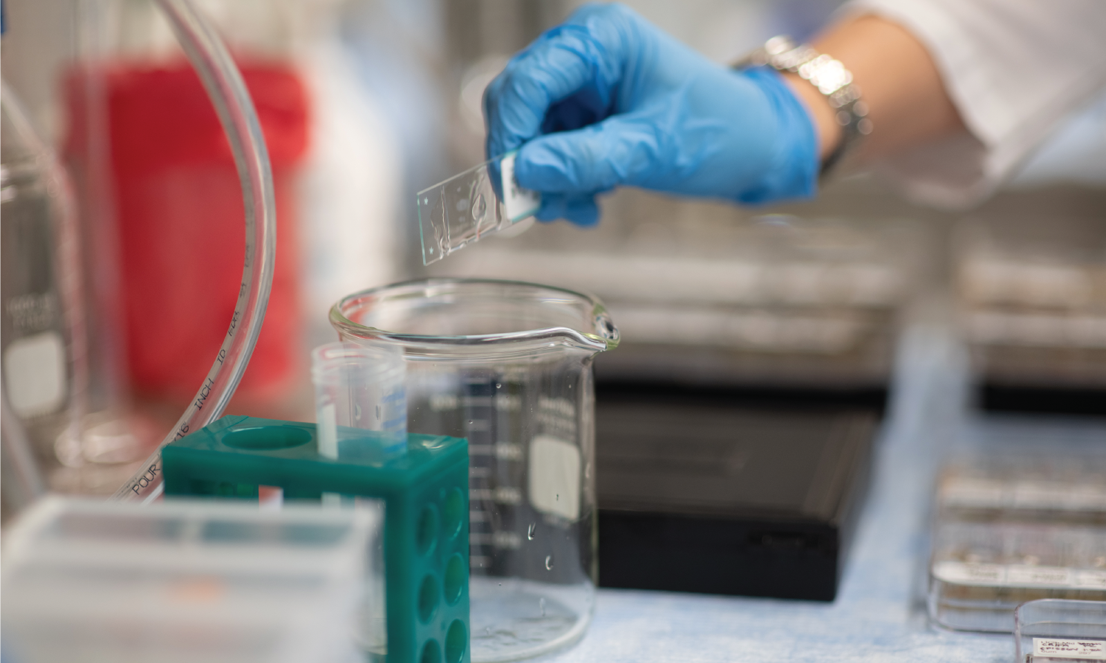
The Core’s knowledgeable staff provide a range of histopathology services, in addition to protocol review and analytical expertise.
Service requests and pricing are managed through Agilent CrossLab (See Schedule Equipment and/or Request Services). Current CrossLab users can login here.
VAI staff that need a login (or further CrossLab support) should contact Lori Moon. MSU and external users who need a CrossLab account/login should go here for account creation. After logging in, external users should click “list all cores” under the Core Facilities section in the left menu and scroll down to “Cores at Van Andel Institute.”
Services
The Core’s pathology services are led by Dr. Galen Hostetter, a board certified pathologist with more than 15 years of experience.
H&E staining is the traditional stain for standard histology and morphology. The use of H&E stained slides to provide specific diagnosis based on morphology is a core skill of anatomic pathology.
IHC staining uses antibodies to detect the abundance, presence, and/or localization of specific proteins in a sample. This staining technique is critical to distinguish between diseases with similar morphology, as well as characterizing the molecular properties of certain cancers.
Antibodies are ordinarily monoclonal (mab), prepared primarily in mouse cells, or polyclonal (pcab), prepared in a variety of living hosts, including rabbit, goat, mouse or donkey, among others.
For multiplex approaches, the Core offers multiple stains that can mark the same or different analytes on the same tissue section. Two different antibodies or a combination of in situ hybridization (ISH) probe (DNA) and an antibody are applied to visualize expression and co-localized proteins at the tissue/cellular level. The ISH probe is used to determine species origin of harvested xenograft tissue. The automated immunostainer platform allows investigator design of additional multiplex strategies, with the potential of up to six markers simultaneously applied to a tissue section. This often requires histotechnician expertise, which the Core provides.
Digital slide scanners create electronic images of an entire microscope slide with superior image quality and speed. These high-quality electronic images can be transferred directly or hosted online for remote review.
Thin sections of tissue are cut on rotary microtomes. Routine thickness for most sections is between 4 and 6 microns.
Fixation alters the tissue by stabilizing the proteins so that they are resistant to degredation. The aim of tissue processing is to remove the water from tissues and replace it with a medium that solidifies the tissue and allows for thin sections to be cut.
The purpose of the tissue microarray (TMA) is to represent (with small samplings of tissue through cores from source blocks) the histomorphological spectrum of the original surgical specimen. Therefore, construction of a TMA that will provide this appropriate representation necessitates careful case choice, case review, array design, technical skill at construction, and quality control. The advantage of a TMA slide is that hundreds of small samples are stained simultaneously on one slide, rather than hundreds of slides, thus saving costs of antibodies, stains, reagents and time.
Molecular Processing
The Pathology and Biorepository Core offers blood and tissue processing, nucleic acid extractions, sample quality assessment, large-scale aliquoting and more. Our highly trained staff will work with you to determine the best method for your exact experimental needs. In addition to our full-service offerings, we also have several pieces of shared equipment available for use.
Service requests and pricing are managed through Agilent CrossLab (See Schedule Equipment and/or Request Services). Current CrossLab users can login here.
VAI staff that need a login (or further CrossLab support) should contact Lori Moon. MSU and external users who need a CrossLab account/login should go here for account creation. After logging in, external users should click “list all cores” under the Core Facilities section in the left menu and scroll down to “Cores at Van Andel Institute.”
Questions? Please email [email protected].
Services
Nucleic acid purifications
- DNA and RNA extractions from tissue, blood, cells, FFPE samples, etc.
- Clean up and concentration of DNA or RNA samples
- Measuring sample concentration and integrity
Blood and frozen tissue processing
- Blood fractionation
- Cell sorting
Sample aliquoting
- Routine aliquoting of fresh blood products (i.e., plasma, buffy coat, whole blood)
- Frozen aliquoting of liquids or tissues using the CryoXtract CXT 353